A Comprehensive Guide to Elbow Arthroscopy

Are you experiencing elbow pain? While this pain can be treated with various non-surgical treatments, sometimes, surgery is the only alternative left to patients.
There is a special type of surgery for resolving elbow joint problems, called arthroscopy. This word is a compound of two Greek words, arthro, meaning joint, and skopein, meaning to look. When translated from Greek, this word means looking within the joint.
The following guide to elbow arthroscopy will introduce you to the procedure.
Anatomy
In order to better understand arthroscopy, you should first gain insight into the elbow anatomy, which is formed by three joining bones, the humerus, the ulna, and the radius. The place where these bones meet is covered with articular cartilage, referring to a protective substance that takes the role of a natural cushion. The rest of the surfaces inside this joint are covered by smooth tissue named synovial membrane.
When an elbow is healthy, the synovial membrane produces enough fluid for lubricating the cartilage and eliminating the friction, which happens when a person rotates his/her arm or bends. Ligaments are present on both the inner and outer sides of the elbow joint to impede dislocation. On its front and backside, the joint is surrounded by muscles. Three nerves cross this surface, which has to be protected during arthroscopy.
This joint enables humans to perform two movements, straightening and bending, also referred to as extension and flexion. These motions occur when the humerus and ulna bone are joined, whereas forearm rotation happens when the ulna and radius join.
When is arthroscopy recommended?
Elbow arthroscopy is recommended when patients suffer from a painful condition, which refuses to respond to nonsurgical treatments, such as medications, physical therapy, and rest. The majority of elbow problems are caused by overuse, injury, and wear and tear. This procedure is generally performed for the removal of loose debris, bone spurs, loosening of the joint capsule, assessing cartilage damage, and tennis elbow. Check out the symptoms, causes, and treatment of cartilage damage.
When suffering from arthritis or an injury, the elbow joint is likely to collect cartilage or loose debris, which results in pain and limited motion. Nevertheless, arthroscopy is a minimally invasive surgery that removes loose debris and eliminates the problem. Additionally, in the first stages of arthritis, bone spurs tend to form near the joint and limit its normal motion. With this surgery, bone spurs can be removed and motion restored.
After experiencing an injury, the elbow joint becomes susceptible to stiffening, which is called arthrofibrosis. The joint capsule can become overly tight, thus limiting the range of motion. In such cases, surgical loosening of the capsule is recommended to prevent the formation of scar tissue following surgery, increasing the chances of regaining motion.
Moreover, arthroscopy is also used for assessing cartilage damage, which cannot be adequately assessed with MRIs and X-rays. After assessing the damage extent, medical professionals decide if additional treatment is needed.
Ultimately, this surgery has proven effective in the treatment of tennis elbow when microscopic tendon tearing happens to the outside of the joint. Although this painful condition can be improved with the help of non-surgical treatments, surgery has proven necessary in some patients. Unlike traditional surgery, arthroscopic surgery allows surgeons to remove the affected tendon without detaching it from the bone. See this URL, https://www.healthline.com/health/fitness-exercise/tennis-elbow-rehab, for some exercises to assist you in the treatment of this condition.
Surgery planning
Prior to this surgery, orthopedic surgeons require a series of evaluations and tests for patients to perform at their primary doctors, such as an electrocardiogram, blood tests, and a chest x-ray. There are supposed to be no health risks for surgeons to perform arthroscopy. Patients should follow the instructions of the doctor before arriving at the hospital.
In addition, patients discuss the anesthesia options with the medical team in charge of anesthesia. This surgery is generally performed with general anesthesia, meaning patients are asleep throughout the procedure. In contrast, regional nerve block injections aren’t usually preferred for this type of procedure, as the numbing effect lasts a couple of more hours after the completion of the procedure. This prevents surgeons from examining the nerves of patients in the recovery room to ensure that the three nerves in this area function well.
Furthermore, these injections are usually administered for pain management. Therefore, if patients need such a regional anesthetic following the nerve examination, it will be provided to them by a member of the medical staff.
Procedure
Once the planning phase is over, the procedure can be performed by administering general anesthesia and intravenous antibiotics. The latter is usually given to patients right before the surgery to minimize the risk of infection following the procedure. Surgeons make sure patients are positioned properly to allow them to adjust their instruments and look inside their elbows. You will be either lying on the side or on your stomach.
When the patient is positioned correctly, the upper arm is put in an arm holder to stay in the right position throughout the procedure. As far as the lower arm is concerned, comprehensive dressing is usually applied to minimize swelling. The skin is then cleaned with antiseptic, after which surgeons start drawing lines on the elbow to mark the nerves, bones, and incision placements.
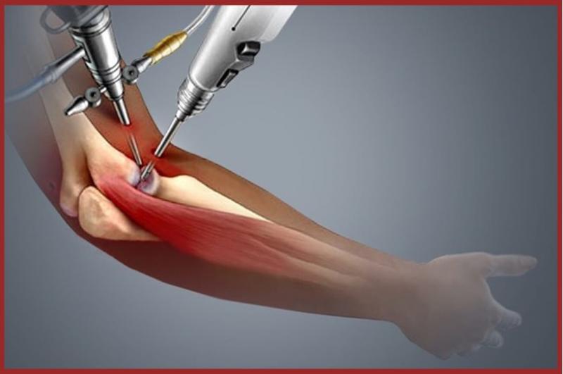
The first thing surgeons do is filling the joint with fluid, which provides them with a clearer view of the structures through the camera. A few tiny incisions are made for the arthroscope to be introduced into the elbow. The images from the instrument are shown on a video screen, providing surgeons with insight into the joint problems.
After performing an evaluation, the doctor starts the required treatment and makes more small incisions if necessary. The incisions are either stitched or covered with a special skin tape once the procedure ends.
The bottom line
Surgery is not always necessary.
Sometimes, however, arthroscopy is the only solution for your elbow joint condition.
It’s minimally invasive, quick, and incredibly effective!
More to Read:


Previous Posts: