What causes TMJ disorders and how do you diagnose them?
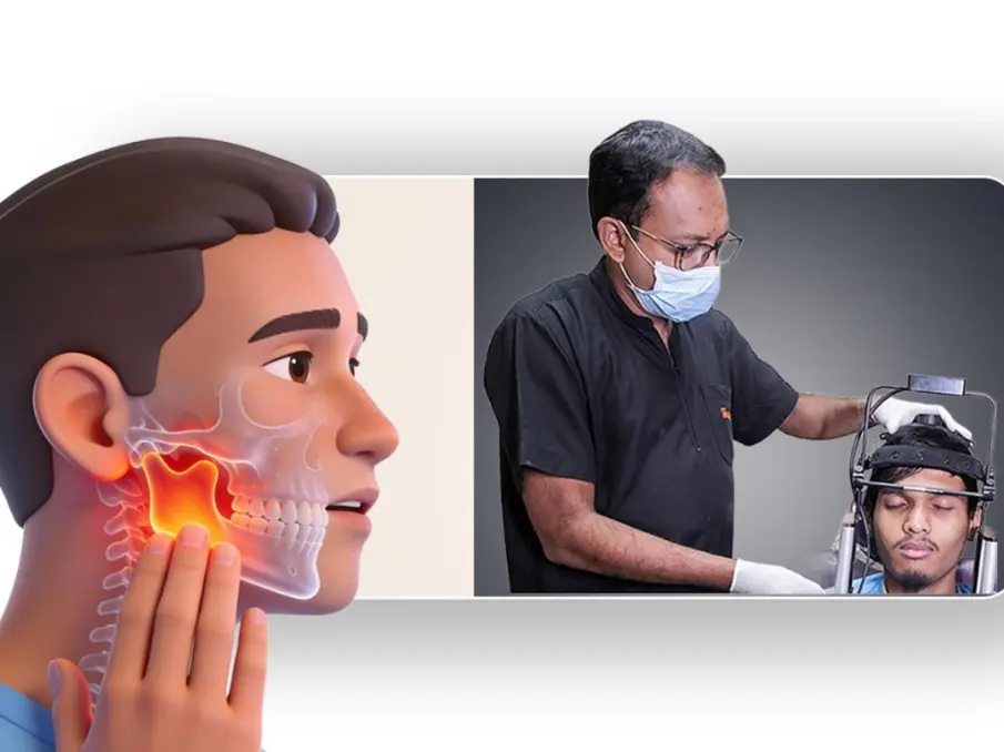
The temporomandibular joint… commonly referred to as the TMJ, is one of the most intricate and heavily used joints in the human body. Located on either side of your face, just in front of the ears, these two joints connect the lower jaw (mandible) to the skull (temporal bone). Every time you chew, speak, swallow, yawn or laugh, these joints perform a complex ballet of hinge-and-sliding motion that most people take entirely for granted… until something goes wrong.
Temporomandibular disorders (TMDs) is the umbrella term for a group of painful conditions affecting this joint, the surrounding muscles of mastication (chewing muscles), and related structures. These disorders affect tens of millions of people worldwide and are among the most common causes of persistent facial pain. Despite their prevalence, TMDs are frequently misunderstood, misdiagnosed or underdiagnosed, because their symptoms can closely mimic those of toothaches, ear infections, migraines and even sinus problems.
Understanding the joint's anatomy
The TMJ is unlike any other joint in the body. Between the rounded head of the mandible (the condyle) and the socket in the temporal bone sits a small but vital structure… the articular disc, a pad of fibrocartilage that absorbs shock and allows smooth movement. The joint is enclosed in a fibrous capsule lined with synovial membrane, which secretes lubricating synovial fluid. Several powerful muscles, particularly the masseter, temporalis and pterygoid muscles, control jaw movement. When any component of this complex system is disrupted, a temporomandibular disorder can develop.
What causes TMJ disorders?
Temporomandibular disorders (TMDs) rarely have a single, isolated cause. In most cases, they arise from a combination of biological, mechanical, psychological and behavioural factors that interact over time. Researchers today generally view TMDs through a biopsychosocial lens… recognising that pain experience is shaped not just by tissue damage but by the nervous system, emotional state, sleep quality and life stressors.
Structural and mechanical causes
One of the most direct mechanical causes is disc displacement… when the articular disc shifts out of its normal position between the condyle and the temporal bone. This is often responsible for the characteristic clicking or popping sound many people hear when opening or closing their mouth. In more severe cases, the disc can become so displaced that the jaw temporarily locks, making it impossible to open the mouth fully.
Arthritis is another significant structural contributor. Both osteoarthritis (the degenerative form common in older adults) and rheumatoid arthritis (an autoimmune condition) can attack the cartilage and bony surfaces of the TMJ, leading to pain, crepitus (a grating sound), and progressive loss of joint function. Less commonly, other inflammatory joint diseases such as psoriatic arthritis or ankylosing spondylitis can involve the TMJ.
Jaw trauma, whether from a direct blow, a motor vehicle accident, a sports injury or even a difficult dental procedure that required prolonged mouth opening, can damage the joint structures, ligaments or disc, precipitating a disorder that may become chronic.
Bruxism and parafunction
Bruxism, the habit of grinding or clenching the teeth, most often during sleep, is one of the most frequently implicated factors in TMD. The forces generated during nocturnal bruxism can far exceed those produced during normal chewing, placing enormous, repetitive strain on the joint and its surrounding muscles. Over time, this can cause muscle inflammation, disc displacement and even bone remodelling within the joint.
Related habits, collectively called parafunctional behaviours, include jaw clenching during waking hours (often unconsciously in response to stress), nail biting, chewing on pens or pencils and habitually resting the jaw in an asymmetric position. These behaviours are particularly problematic because they occur in the absence of any protective proprioceptive feedback, the brain's normal mechanism for regulating bite force is largely bypassed.
Malocclusion and dental factors
The relationship between bite (occlusion) and TMD is an area of ongoing debate among clinicians. Significant malocclusions, such as a severely open bite, crossbite or the absence of posterior teeth, can place asymmetric loads on the joint over time. Poorly fitting dental restorations, dentures or orthodontic appliances that alter the bite plane can also trigger or worsen symptoms. However, it is important to note that many people with obvious malocclusions never develop TMD, suggesting that occlusal factors alone are rarely sufficient.
Psychological and psychosocial factors
Stress, anxiety and depression have a well-established relationship with TMDs. Psychological stress commonly manifests as muscle tension, particularly in the jaw, neck and shoulder muscles. This tension can become chronic, contributing to the myofascial pain component of TMD. People with depression and anxiety also tend to report higher pain severity and are at greater risk for the condition becoming chronic and disabling.
Sleep disturbances frequently co-occur with TMDs and the relationship appears bidirectional… poor sleep increases pain sensitivity and facial pain disrupts sleep quality, creating a self-perpetuating cycle.
Hormonal influences
TMDs are significantly more common in women of reproductive age than in men or post-menopausal women, suggesting that oestrogen may play a role in joint pain sensitivity and inflammation. Some research points to oestrogen receptors in TMJ tissues, raising the possibility that hormonal fluctuations influence disc health and pain thresholds. This is an active area of investigation and may eventually influence treatment strategies.
Hypermobility and connective tissue disorders
Individuals with generalised joint hypermobility and especially those with connective tissue disorders such as Ehlers-Danlos syndrome are at heightened risk for TMDs. The lax ligaments that characterise these conditions provide insufficient support to the TMJ, allowing excessive translation of the condyle and predisposing the disc to displacement.
Neurological and central sensitisation factors
In chronic TMDs, central sensitisation… a state in which the central nervous system becomes hyper responsive to pain signals, plays a critical role. This explains why some patients continue to experience significant pain even after the original tissue injury has resolved, and why their pain may spread beyond the jaw to the neck, head, and elsewhere. The overlap between TMD and other chronic pain conditions such as fibromyalgia, irritable bowel syndrome, and chronic fatigue syndrome is striking and reflects this shared neurological mechanism.
How is TMD diagnosed?
Diagnosing Temporomandibular disorders (TMDs) requires careful integration of clinical history, physical examination and when indicated - imaging. There is no single definitive test and diagnosis is primarily clinical. The most widely used research and clinical diagnostic framework today is the Diagnostic Criteria for Temporomandibular Disorders (DC/TMD), which provides validated, standardised criteria for classifying different subtypes of TMD.
Clinical history
A thorough history is the cornerstone of diagnosis. The TMJ Specialist, typically an oral medicine and maxillofacial radiologist, a dentist with TMD training will enquire in detail about the nature, location, duration and triggers of pain. Key questions include whether pain occurs at rest or only with jaw movement, whether it is unilateral or bilateral, what makes it better or worse, whether there are associated symptoms such as headache, ear pain, tinnitus or dizziness, and whether there is a history of trauma, stress or sleep problems.
Physical examination
The physical examination includes palpation of the masticatory muscles, the masseter, temporalis and pterygoid muscles, to identify tenderness and trigger points. The clinician will also palpate the joint itself, both laterally and via the external auditory meatus (ear canal), to detect tenderness or crepitus directly over the condyle.
Jaw range of motion is carefully measured. Normal maximum mouth opening in adults is typically 40–55 millimetres between the upper and lower incisors. Restricted opening (under 40 mm) may indicate disc displacement without reduction, muscle splinting or intra-articular pathology. Deviations or deflections of the jaw during opening, where the jaw swings to one side can point to unilateral disc involvement. The clinician will also assess lateral excursions and protrusive movements.
Joint sounds are documented. Clicking early in the opening cycle (early opening click) combined with a click on closing (reciprocal click) is characteristic of anterior disc displacement with reduction. The absence of clicking in a jaw that was previously clicking, combined with suddenly reduced opening, suggests progression to disc displacement without reduction… a locked jaw.
Questionnaires and symptom scales
The DC/TMD protocol incorporates validated self-report tools including the Patient Health Questionnaire (PHQ-4) for screening depression and anxiety, the Jaw Functional Limitation Scale and pain drawing maps. These psychosocial assessments are not merely supplementary in chronic TMD, they are central to understanding the full picture and guiding treatment.
Imaging
Imaging is not required for every patient but becomes important when the diagnosis is uncertain, when structural pathology is suspected or when surgical management is being considered.
Panoramic radiography (OPG) is often the first-line imaging study, providing an overview of the TMJs, teeth and jaw. It can reveal obvious bony changes such as condylar erosion, flattening or osteophyte formation, but it provides limited detail and cannot visualise soft tissues.
3D Cone beam computed tomography (CBCT) offers far superior bony detail and is the preferred modality for assessing condylar morphology, articular surface changes and bony ankylosis. CBCT has become increasingly accessible in dental practices and delivers a relatively low radiation dose compared to conventional CT.
Magnetic resonance imaging (MRI) is the gold standard for visualising the articular disc and surrounding soft tissues. It can precisely determine disc position, shape and morphology, identify joint effusion (fluid accumulation within the joint space, a sign of active inflammation) and detect adjacent soft tissue abnormalities. MRI is the investigation of choice when disc displacement, inflammatory arthritis or retrodiscal tissue changes are suspected.
Ultrasound has emerged as a useful dynamic, real-time imaging tool that can visualise the disc and detect joint effusion without radiation. It is operator-dependent and less widely used, but holds promise as an accessible, cost-effective adjunct.
Blood examination and nutritional assessment
Blood tests help rule out systemic conditions that mimic or worsen TMD. Inflammatory markers such as ESR and CRP can point to arthritis affecting the joint, while nutritional panels check for deficiencies in vitamin D, calcium and magnesium… all of which influence bone integrity, muscle tension, and pain sensitivity. Thyroid function is also assessed, as hypothyroidism can manifest as generalised jaw and muscle stiffness.
T-Scan — digital occlusal analysis
T-Scan is the only clinically validated digital system for measuring bite force distribution in real time. Using a thin sensor wafer, it maps exactly where, how hard, and in what sequence teeth make contact… identifying premature contacts and left-right force imbalances that create asymmetric loading on the TMJ. It takes the guesswork out of occlusal balancing entirely.
BioPAK diagnostic system
BioPAK is an integrated platform that combines jaw tracking, electromyography, and joint vibration analysis in a single coordinated system. Its greatest strength is the ability to overlay all three data streams simultaneously… revealing, for example, the exact moment in the jaw's movement arc that a muscle fires abnormally, correlating structure and function in one view.
3D jaw tracker
The JT-3D tracks mandibular movement with millimetre precision across all three planes of space… vertical, horizontal and sagittal. It quantifies opening range, movement speed, lateral deviations, and the true three-dimensional rest position of the jaw. This data is essential for identifying condylar restriction, planning orthotic therapy, and objectively monitoring functional improvement over time.
Electromyography (EMG)
Surface EMG records the electrical activity of the masticatory muscles… primarily the masseter and temporalis. In healthy function, these muscles fire symmetrically and show minimal resting activity. TMD patients typically show elevated resting tension, left-right asymmetry, and abnormal firing patterns during chewing. EMG provides objective, measurable evidence of muscle dysfunction before and after treatment.
BioJVA — joint vibration analysis
BioJVA captures the vibrations produced by both TMJs simultaneously as the patient opens and closes their mouth. Low-frequency vibrations indicate disc displacement; high-frequency patterns point to degenerative bony changes. It produces a graphically documented, reproducible record of intra-articular pathology… far more objective and specific than listening with a stethoscope, and with no radiation involved.
Airway volumetry
Using CBCT data processed through 3D software, airway volumetry calculates the precise volume and narrowest cross-section of the upper airway. Since a retruded mandible directly reduces posterior airway space, this assessment reveals whether jaw repositioning could simultaneously relieve joint loading and improve breathing… linking TMD treatment directly with airway and sleep medicine.
Polysomnography (sleep study)
A full-night sleep study records brain activity, oxygen saturation, respiratory airflow, and crucially, masticatory muscle activity via EMG… the electrophysiological signature of sleep bruxism. It determines whether nocturnal grinding is actually occurring, how severe it is, and whether obstructive sleep apnoea is the underlying trigger. For patients where sleep is driving their TMD, polysomnography is indispensable for shaping the right treatment plan.
Diagnostic anaesthetic injections
In cases where the source of pain is ambiguous; for example, when it is unclear whether pain originates from the joint itself or from the surrounding muscles, a diagnostic injection of local anaesthetic directly into the joint space or a specific muscle can help localise the pain source. Temporary elimination of pain following injection confirms that structure as the pain generator.
Why early and accurate diagnosis matters?
Because TMDs exist on a spectrum from mild and self-limiting to severe and disabling, early and accurate diagnosis enables appropriate stratification of care. Many patients respond well to conservative, reversible treatments, patient education, jaw rest, dietary modification, physiotherapy, occlusal splints and psychological support. A minority with significant structural pathology or refractory pain may require more specialised interventions. Misdiagnosis… particularly mistaking TMD for an ear infection, dental abscess or primary headache disorder, delays effective treatment and can allow acute problems to transition into chronic pain states.
At FMS International Dental Center, patients have access to one of the most comprehensively equipped TMD diagnostic setups available anywhere. Since 2018, FMS TMD Clinic has housed the full spectrum of advanced diagnostic technologies described in this article, from T-Scan digital occlusal analysis and BioJVA joint vibration analysis, to 3D jaw tracking, surface EMG, airway volumetry and in-house polysomnography coordination, all under one roof. For more than eight years, a dedicated team of senior TMJ specialists at FMS has been using this integrated suite of tools daily, bringing a depth of clinical experience that is rare even by international standards. Every patient who walks in with jaw pain, clicking, facial discomfort or suspected TMD is assessed not with guesswork, but with objective, reproducible, multi-dimensional data, ensuring that the true root cause is identified and that treatment is precisely targeted from day one.
More to Read:








Previous Posts:

